Our reception areas are comfortable and stocked with attractive items for your pets. As well as our four consulting rooms, we have modern glass-fronted kennels for pets spending the day with us, an x-ray suite and prep room, a fully equipped modern operating theatre and a pharmacy room. Our dental units provide facilities for all routine veterinary dentistry procedures. Diagnostic and surgical facilities include x-ray, dental x-ray, ultrasound, electrocardiography, Doppler blood pressure, and a range of diagnostic endoscopes. These are explained in more detail below.
Internal Medicine
Internal medicine services cover hormonal, gastrointestinal, urinary, haematologic (blood related), respiratory, infectious, and immune-mediated diseases. The facilities at the London Veterinary Clinic are co-ordinated by Grant Petrie. In addition to an extensive review of your pet’s medical history, diagnostics always begin with a thorough physical examination and blood tests. When more information is needed, ultrasound, radiography, endoscopy, and CT imaging may be used. Our objective is to make an accurate diagnosis as rapidly as possible while at the same time putting your pet (and you) to a minimum of inconvenience.
Cardiology
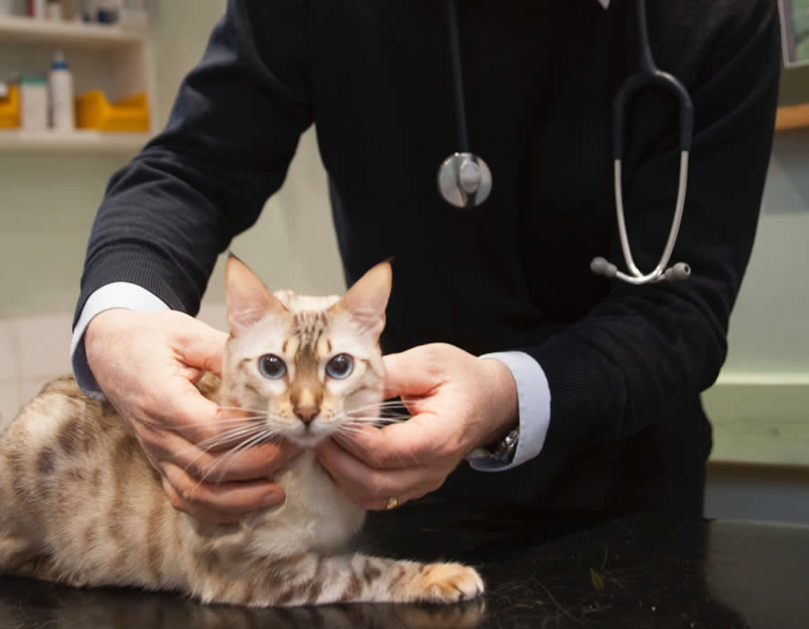
Cardiology services are provided by Grant Petrie. In addition to an extensive medical history review, a consultation may include echocardiography, electrocardiography, radiography, blood pressure monitoring and 24 hour Holter monitoring to evaluate the cardiac status of your pet.
Behaviour
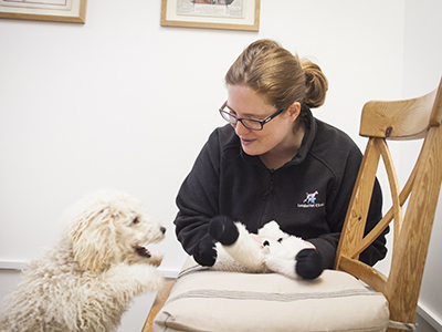
Our behaviour service at London Vet Clinic is provided by veterinary behaviourist, Natalia Aira Bewick. Our bespoke service can help you manage and prevent any behaviour problems in dogs and cats.
A thorough, individual assessment is carried out to provide you with a tailored behaviour therapy plan, this may include a training plan, medical diagnostics, behaviour medications (psychotropics) and/or supplements. Natalia has also created a unique puppy course exclusive to the London Vet Clinic.
Dentistry
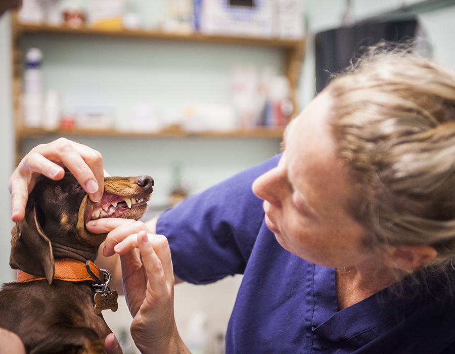
Preventing tooth and gum disease is vital and when you visit us we explain how to keep your companion’s mouth, teeth and gums healthy. The reality is, however, that oral problems are common and because they are we are experienced in and have the sophisticated equipment needed to provide successful care. Dental x-rays are particularly important to help reveal tooth damage below the gum line. Dental extractions when necessary, scaling and polishing, are all time consuming procedures. There are usually two vet nurses assisting the vet with each dental.
Occasionally unusual dental procedures are needed and when they are these can be done at York Street, without the need for travel to a referral hospital by our dentist Peter Kertesz who has over 40 years of experienced in animal dentistry (and who wrote the first textbook on comparative dentistry).
Dermatology
We will take a full medical history and may take an ear or skin swab or skin scrapings to help with the diagnosis. In rare and unusual skin conditions a skin biopsy may be needed. For allergic skin conditions exclusion diets are often recommended and allergy testing for inhaled allergies (pollens, moulds, dust mites etc) is done on blood samples sent to a specialist laboratory in The Netherlands. We have specialist referral veterinary dermatologist available for the most challenging cases.
Oncology
Cancer is a common condition in older dogs and cats. Recent advances in the treatment of cancer in pets have opened up a number of options (and also ethical dilemmas) for pet owners and your pets. Even with these advances however, surgery remains an essential component of treatment for most types of cancer.
Our aim at the London Veterinary Clinic is to extend an enjoyable and pain-free life for pets. Our vets undertake surgery and chemotherapy. When radiotherapy is appropriate it is undertaken at Cambridge University. Making the decision to pursue surgery, radiotherapy or chemotherapy for the treatment of a cancer can be difficult and stressful – particularly when we have personal experience with cancer in our own families. It raises the ethical question of how far we should go in treating our pets’ medical conditions.
Our judgment and skill can help you understand your pet’s problem, and whether surgery cures or contains the condition.
Ophthalmology
An initial consultation includes a history taking and eye examination. We have the same equipment for measuring the pressure in the eyes (glaucoma) as you may have experienced when visiting an optician or ophthalmologist. As well as ophthaloscopes in all the exam rooms we also have an advanced slit lap for detailed examination of all parts of the eyes. Our GP vets are experienced with treating all common eye conditions but when they feel that it’s in your pet’s interest to be seen by a specialist, Dr David Williams visits York Street. Dr Williams is Cambridge University’s vet school’s resident ophthalmologist. When eyes need surgical repairs these can be done at York Street and your companion is back home the same day.

Diagnostic Imaging
Among the most useful tools we use to evaluate a pet’s condition is diagnostic imaging. This includes radiographs (x-rays) and ultrasonography (ultrasound) both of which are carried out at York Street.
We are able to refer pets for other forms of imaging if needed, this may include computed tomography (CT scans) and magnetic resonance imaging (MRI scans). Each of these provides different kinds of images, and in some instances more than one may be suggested by us to evaluate a particular problem in your pet. All of these methods provide ‘pictures’ of internal structures.
Read More
X-rays
The most familiar imaging method and most frequently used at the London Veterinary Clinic is radiography or x-rays. Anatomy can be discerned because of differences in contrast between fat, water, bone and metal. Most of the body’s soft tissues are either water-dense or fat-dense. The most commonly taken x-rays are those of the chest cavity, abdominal cavity, limbs, spinal column, and skull.
Because dogs and cats are generally unwilling to hold still, especially in an awkward position, some form of restraint is usually required to take x-rays. While being held by people is a simple form of restraint, hand-restrained pets may still squirm. We often have to take more than a single film and this adds to the potential distress for the dog or cat and potential radiation exposure for people holding the pet. While the amount of radiation received by the pet is minimal, even with multiple x-rays, our concern is cumulative dosing to people. This is why we use alternative methods of restraint.
Where appropriate we keep a fully conscious pet still with well placed soft-covered surrounding sandbags. There are instances where this is inadequate so we selectively use various sedatives or anesthetics. There are many choices of drugs and often only a brief sedation from which a pet is fully recovered within just a few minutes is used. We will discuss with you the need for x-rays and what type of restraint is needed.
Contrast x-rays
In addition to plain x-rays we sometimes use radiographs to do contrast studies. In these instances a dye material is administered that on radiographs has a ‘metallic’ density. The contrast agent most of us are familiar with is barium, and this is sometimes used to evaluate the digestive tract. However, contrast studies are also used with different dye agents to evaluate the urinary system (excretory urograms or intravenous pyelograms – IVP’s), blood vessels (angiograms), and spinal cord (myelograms).
Ultrasound
The next most commonly used imaging method at the London Veterinary Clinic is ultrasonography. High frequency sound waves are used to provide more narrowly focused pictures of internal structures. Details that might be difficult to see on radiographs can often be seen with ultrasound. Ultrasound can also provide “real-time” images of structures to detect such things as movement. Most of us are familiar with ultrasonography to ‘image’ developing fetuses in pregnant women. Real time imaging also permits ultrasound-guided aspiration (placing a small needle into a structure to collect some fluid or cells as is done for amniocentesis in some human pregnancies) or biopsy of internal structures.
More so than radiographs, the success of ultrasonic imaging is directly linked to the skill and training of the ultrasonographer. Individuals highly trained in ultrasound imaging may be able to make diagnoses, obtain samples, and provide treatment advice far better than less skilled or experienced vets even when using identical machines. Grant Petrie and Veronica Aksmanovic undertake our ultrasonography.
Computed tomography uses x-rays in a 360-degree technique with a special scanning machine. Like plain radiographs the amount of radiation exposure for patients with CT is minimal. Technology has progressed dramatically. Current CT scanners allow complete imaging of even large areas in just a few minutes, and the digital images can then be reformatted to provide 3-dimensional reconstructions. Because patient motion greatly degrades the images, pets are always given short-acting sedation/anesthesia for CT scans. We refer pets for CT scans to our associates at The Ralph Vet Referrals and Dick White Referrals.
Magnetic resonance imaging (MRI) is a non-x-ray based technology. It uses a powerful magnet and radiofrequency waves to create highly detailed images of virtually any structure in the body. MRI pictures can provide far more detail than either x-rays or ultrasound. All MR imaging in animals requires anesthesia due to the need for complete immobility during scanning and the relatively longer time needed for MRI than other imaging techniques such as x-rays or CT. There is some overlap between the indications for CT and MRI scanning. The former is generally best for bony details such as elbows, knees and complex fractures. CT is also excellent for pictures of the lungs, and conditions in the ears and nose. MRI is generally superior for brain, spinal cord, and abdominal imaging. If we recommend advanced imaging we will discuss with you the reasons for our preference for either CT or MRI. MRI scans are conducted by our colleagues at The Ralph Veterinary Referrals and Dick White Referrals.
Fluorsocopy is used on occasion to evaluate such things as swallowing function, angiography (motion studies of blood flow or interventional radiology and interventional cardiology using special catheters), and can be used in orthopedic surgery to evaluate placement of drills, screws, and so forth. It is real-time x-ray imaging. Fluoroscopy is used to place cardiac pacemakers and vessel stents or coils. When needed, fluoroscopy is undertaken at Cambridge University.
Scintigraphy involves the administration of minute quantities of a radioactively-labeled substance into the body, then monitoring and measuring where the radio-labeled substance goes after administration. Bone scans are one form of scintigraphy. Thyroid scans are another. The pictures provided with scintigraphy are crude compared with other imaging modalities, but have the advantage of providing functional images, indicating normal or abnormal function of target organs. We refer patients to Cambridge University for scintigraphic evaluations.
Hospitalisation
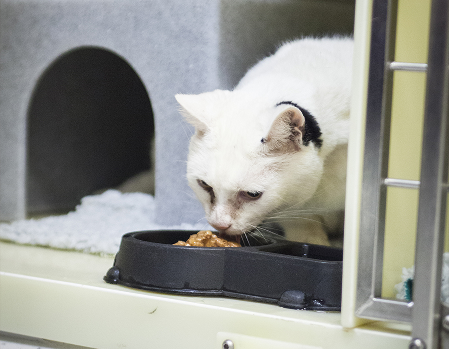
At the London Vet Clinic, we undertake all procedures as ‘out-patient’ activities. He have glass fronted day ‘dens’ for each dog or cat as well as larger walk-in facilities for giant dogs. They are set up so that no animal is looked at by another. Animals needing extra oxygen have their own oxygen ‘den’.
When a dog or cat needs continuing care overnight we will arrange for this to be done at a 24 hour facility, usually the Medivet out-of-hours facility in Kensington.
Laboratory
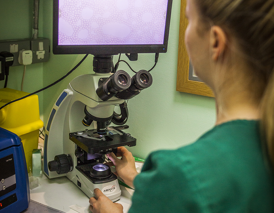
Our Lab facilities at the London Vet Clinic are provided, calibrated and maintained by Idexx, the world’s largest provider of veterinary lab diagnostics. We undertake routine haematology and biochemistry test but also hormone and antibody assays and urine analyses. Our digital screen microscope means that several vets can review samples. We routinely do microscope exams of ear discharges to find the specific cause of these problems.
In additional to our extensive York Street lab facilities we send samples to off-site labs such as the University of Glasgow and the University of Bristol for specialist tests.


